

“It never hurts your eyesight to look on the bright side of things.” Barbara Johnson
“Vision is seeing visibly the light of hope within the range of the eyesight.” Anuj
Introduction: There is no doubt that with age comes a worsening of vision. Furthermore, there is also an effect on one’s eyesight by Parkinson’s. Consider the following scenario. A person-with-Parkinson’s (PwP) walks with friends early in the evening, and it is cloudy outside as the early evening approaches. The PwP realizes that their sharpness of sight has changed, and suddenly, they fall and fracture their hip. What caused the fall of the PwP? Likely, the combination of posture and gait issues combined with changes in visual acuity. Herein is a review of the many different problems one can obtain regarding vision in the presence of Parkinson’s.
“There is nothing invisible in this universe! There is only our lack of eyesight!” Mehmet Murat Ildan
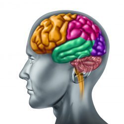
Dopamine: You already know the answer, but what substance is depleted to cause Parkinson’s? Yes, it’s dopamine, which is a critical neurotransmitter (or chemical messenger). Most of our dopamine is made/synthesized in the mid-brain region by dopaminergic neurons in the substantia nigra pars compacta region and the ventral tegmental area. There are five different dopamine pathways to support or store dopamine for various physiological functions. They include Nigrostriatal, Mesocortical, Mesolimbic, Tuberoinfundibular, and Retinal Fibers. Dopamine combines with serotonin, acetylcholine, norepinephrine, and other neurotransmitters to orchestrate many essential physiological functions. The drawing below highlights the role of dopamine in the brain.

Dopamine helps us stay focused and allows us to remain attentive. Vision can help with the dopamine response in the brain that then assists us in focusing. Parkinson’s begins because the dopaminergic neurons stop producing dopamine, which starts a cascade of events leading to both motor- and non-motor dysfunctions. The aspects here in this post dealing with vision are considered non-motor-related problems.
“No person ever ended his eyesight by looking on the bright side.” Zig Ziglar
The Eye, Retina, and Dopaminergic Neurons: The goal here is not to fully describe the eye and vision but to present the retina’s function regarding sight and how dopamine functions in this critical region.
Think of the retina as a transformer that converts the light that enters the eye into electrical signals. These signals allow the optic nerve to signal the brain that generates the images one sees.
Dopamine in the retina modulates light adaptation. Amacrine cells are the intrinsic neurons of the inner retina. These cells release dopamine when activated. As described in the Encyclopedia of the Eye (click here): “Amacrine cells (ACs) are multipolar retinal neurons branching within the inner plexiform layer of the retina to collect and decode bipolar cell (BC) signals, recoding them as synaptic release patterns of 4-aminobutyrate (gamma aminobutyric acid), glycine, and other neurotransmitters to modulate the activity of ganglion cells (GCs).” This passage says that dopamine is a chemical messenger for light adaptation by promoting the flow of information from cone circuits while diminishing rod circuits.

The diagram from Wikipedia (attribute here) gives the structure of the eye.[Rhcastilhos. And Jmarchn., CC BY-SA 3.0 https://creativecommons.org/licenses/by-sa/3.0, via Wikimedia Commons]
“Eyes that look are common; eyes that see are rare.” J. Oswald Sanders
Parkinson’s and Vision Changes: It has been estimated that visual symptoms are present in >75% of People-with-Parkinson’s (PwP). What typically is found are changes in visual acuity, spatial contrast sensitivity, and color vision.
I can read the computer screen more easily without my new bifocal glasses. Furthermore, gauging distances at night has become more challenging when observing cars and trucks come and go past me. It is unclear whether these two aspects of my vision relate to me just getting older or are linked to a dopamine deficiency caused by Parkinson’s.
I do not ‘see the light’ better when levodopa is being converted to dopamine in my brain following any daily dose of carbidopa/levodopa, in contrast to the improved motor function I can identify during such treatment. However, that could be a supply and demand issue between my intake of carbidopa/levodopa, the processing in the brain, and the dissemination of this chemical messenger to the appropriate site for action. However, several papers suggest that levodopa medication can improve certain eye disorders.
I will also stop blinking sometimes, and Susan must remind me to blink! This not-blinking phenomenon is especially apparent when concentrating while I am driving.
“The only thing worse than being blind is having sight but no vision.” Helen Keller’
Clinical Conditions Associated with Vision Changes from Parkinson’s: The chart below lists some of the more common changes that can occur in your eyes in Parkinson’s.
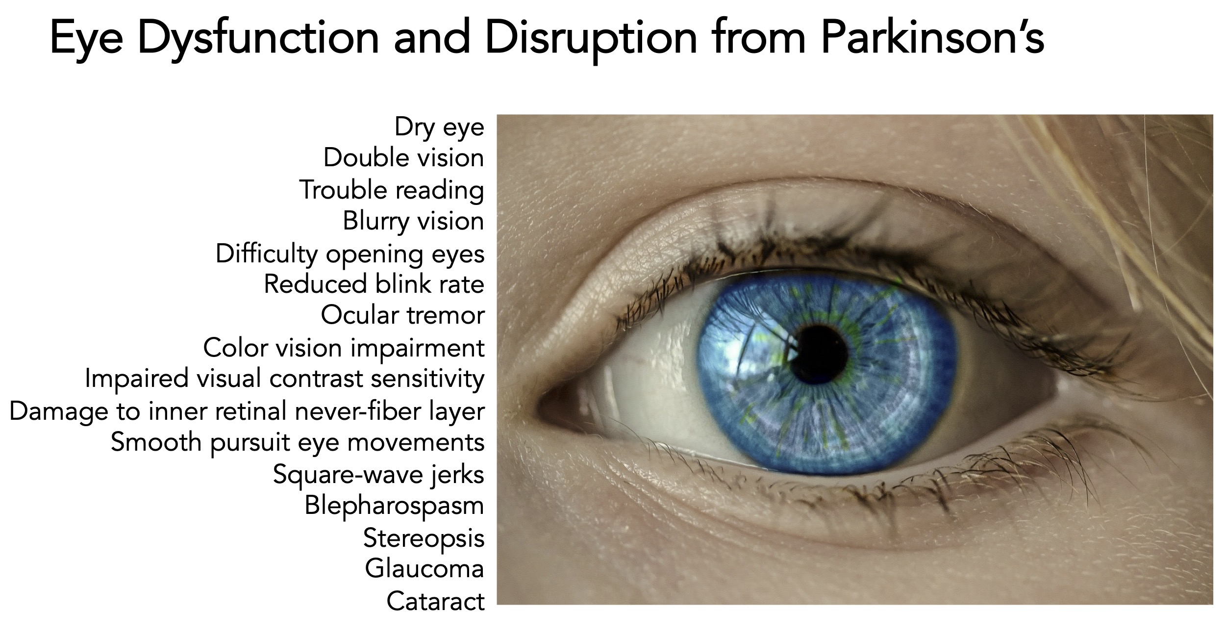
•Dry eye- Dry eye syndrome, with reduced eye secretion, is quite common. Using artificial tears and good eyelid cleanliness will help.
•Double vision- Diplopia (or double vision) causes people to see two of the same image—whether horizontal, vertical, or diagonal—instead of one.
•Trouble reading– A PwP may have trouble reading due to vision changes, making it harder to easily discern words on a page, or it could be linked to cognitive changes in the PwP.
•Blurry vision- The inability to focus your eyes, as mentioned above, may lead to blurry vision.
•Difficulty opening eyes– Apraxia of eyelid opening (ALO) is a non-paralytic inability to open the eyelids purposedly. Difficulty opening your eyes is linked to levels of levodopa in the patient.
•Reduced blink rate- This is likely associated with dopamine levels, and blink rate may reflect striatal and mesolimbic dopaminergic activity.
•Ocular tremor- Like tremors in the hands, arms, legs, etc., a PwP may also express an ocular tremor.
•Color vision impairment– Some PwP exhibit impaired vision when tested for color thresholds and contrast sensitivity.
•Impaired visual contrast sensitivity– Poor visual contrast sensitivity does not affect the ability to see objects, except when these objects are the same or similar colors as their surroundings.
•Damage to inner retinal never-fiber layer– One study found the inner retinal layers suffered more obvious thinning in PwP with long disease duration, which diminishes vision.
•Smooth pursuit eye exercises (SPEM)- SPEM are conjugate eye movements performed to keep a moving object fixated on the fovea (a slight depression in the eye’s retina where visual acuity is highest). SPEM in PwP has been evaluated for accuracy, speed, and quality. SPEM abnormalities are more significant in the Parkinson’s population, with an estimated prevalence of 67% in Parkinson’s compared to 20% in healthy age-matched controls.
•Square-wave jerks– Square-wave jerks are saccadic (a saccade is a rapid eye movement that shifts the center of gaze from one part of the visual field to another) intrusions (usually 0.5–5°) that move the eye from and back to the fixation point with an intersaccadic interval of about 200 ms. Eye movements in the saccadic system allow one to test a network involving cortical (mainly frontal and parietal) and subcortical (basal ganglia, midbrain, brain stem, thalamus, and cerebellum) structures.
•Blepharospasms- Blepharospasms in PwP are of great concern because they obstruct the patients’ visual field. Blepharospasm is an involuntary spasm of the orbicularis oculi muscle and is considered focal dystonia. It is more prevalent in atypical parkinsonism.
•Stereopsis- Depth perception problems exist in PwP. Disruption due to stereopsis gives one difficulty navigating an automobile and makes the PwP more susceptible to falls.
•Glaucoma– PwP have an increased risk of developing glaucoma. It was shown that glaucoma patients have reduced levels of dopamine and other neurotransmitters, which suggests that the dopaminergic action is altered in the eye. Thus, the reason for increased glaucoma risk in Parkinson’s.
•Cataract– PwP are also more likely to develop cataracts when compared to the average population. Lens removed from PwP with cataracts had increased alpha-synuclein deposits compared to normal controls. This suggests that Parkinson’s and the lens that form cataracts potentially share a common dopaminergic-linked pathway.
“My eyesight’s gone, my reflexes are shot, and I can’t stay awake, but thank God I can still drive.” Robert Breault
Vision Changes, Parkinson’s, and Quality-of-Life: No doubt, vision changes as we age. As described here, numerous changes to our eyes are modified by Parkinson’s. These vision problems can contribute to a reduced quality-of-life in Parkinson’s. Furthermore, PwP read fewer words per minute than age-matched controls.
As expected, vision difficulty, postural instability, and gait problems might increase the chance of falling.
Unfortunately, as Parkinson’s progresses, visual impairment is realized. Color vision, stereopsis, and contrast sensitivity changes are linked to disorder progression. Ultimately, the conclusion reached from many studies is that vision issues are linked closely to the decline of dopaminergic neurons inside the retina.
“How alive is thought, invisible, yet without thought there is no sight. Dejan Stojanovic
Assessment of Vision Issues in Parkinson’s: I admit that learning about the various vision issues in Parkinson’s and then writing this blog post startled me somewhat. I was aware of dopaminergic neurons in the retina, but the variety of vision issues found in Parkinson’s was unexpected. Review some of the papers included below. They are all quite good reading and very educational.
If you are experiencing vision problems, please look through the paper by Borm et al. (Borm, Carlijn DJM, Katarzyna Smilowska, Nienke M. de Vries, Bastiaan R. Bloem, and Thomas Theelen. “The neuro-ophthalmological assessment in Parkinson’s disease.” Journal of Parkinson’s disease 9, no. 2 (2019): 427-435.); it has a comprehensive overview of how a neuro-ophthalmological assessment should be performed.
“The eye sees only what the mind is prepared to comprehend.” Robertson Davies
Concluding Thoughts: The overall goal of this blog post was to increase awareness about the complex vision issues of Parkinson’s. Understanding that simple problems like dry eye syndrome can be treated. Vision changes in Parkinson’s may require multiple glasses to properly visualize everything. More difficult vision problems occur in Parkinson’s and require your vigilance and expert medical advice during the journey. In conclusion, the eyes have it. They are affected by Parkinson’s. Continue good health to all, and continue reading, and keep challenging your cognition.
“Small is the number of them that see with their own eyes and feel with their own hearts.” Albert Einstein
Useful References:
Archibald, Neil K., Michael P. Clarke, Urs P. Mosimann, and David J. Burn. “The retina in Parkinson’s disease.” Brain 132, no. 5 (2009): 1128-1145.
Di Pippo, Mariachiara, Serena Fragiotta, Federico Di Staso, Luca Scuderi, and Solmaz Abdolrahimzadeh. “The role of alpha-synuclein deposits in Parkinson’s disease: a focus on the human retina.” International Journal of Molecular Sciences 24, no. 5 (2023): 4391.
Zhang, Yanyan, Xiaoguang Zhang, Yunhua Yue, and Tian Tian. “Retinal degeneration: a window to understand the origin and progression of Parkinson’s disease?.” Frontiers in Neuroscience 15 (2022): 799526.
Ba, Fang, Tina T. Sang, Wenjing He, Jaleh Fatehi, Emanuel Mostofi, and Bin Zheng. “Stereopsis and eye movement abnormalities in Parkinson’s disease and their clinical implications.” Frontiers in Aging Neuroscience 14 (2022): 783773.
Stuparu, Alina Zorina, Sanda Jurja, Alexandru Floris Stuparu, and Any Axelerad. “Narrative Review Concerning the Clinical Spectrum of Ophthalmological Impairments in Parkinson’s Disease.” Neurology International 15, no. 1 (2023): 140-161.
Borm, Carlijn DJM, Katarzyna Smilowska, Nienke M. de Vries, Bastiaan R. Bloem, and Thomas Theelen. “The neuro-ophthalmological assessment in Parkinson’s disease.” Journal of Parkinson’s disease 9, no. 2 (2019): 427-435.
Chorostecki, Jessica, Navid Seraji-Bozorgzad, Aashka Shah, Fen Bao, Ginny Bao, Edwin George, Veronica Gorden et al. “Characterization of retinal architecture in Parkinson’s disease.” Journal of the neurological sciences 355, no. 1-2 (2015): 44-48.
Harnois, Carmen, and T. Di Paolo. “Decreased dopamine in the retinas of patients with Parkinson’s disease.” Investigative ophthalmology & visual science 31, no. 11 (1990): 2473-2475.
Ortuño‐Lizarán, Isabel, Thomas G. Beach, Geidy E. Serrano, Douglas G. Walker, Charles H. Adler, and Nicolás Cuenca. “Phosphorylated α‐synuclein in the retina is a biomarker of Parkinson’s disease pathology severity.” Movement Disorders 33, no. 8 (2018): 1315-1324.
“Eastward the dawn rose, ridge behind ridge into the morning, and vanished out of eyesight into guess; it was no more than a glimmer blending with the hem of the sky, but it spoke to them, out of the memory and old tales, of the high and distant mountains.” J. R. R. Tolkien
Cover Photo Image by Paul Brennan from Pixabay

My Dad’s difficulties reading a book centered around the boo moving, until he built a little holding stand.
LikeLiked by 1 person
Makes total sense to secure the book in a stand.
LikeLike
My PWP Dad suffered eyelid apraxia. He was a huge reader and it was such a frustration for him. Before that he’d had quite strong prisms added to his myopia prescription. In the earlier days, he’d be able to do things like stand up to make his eyes pop open. Eventually it impaired his driving — took the keys away the day his eyes (that he said always used to open when the car moved) didn’t open and he backed over a retaining wall.
The saddest part was when he was in the hospital for a fall and the aide came in and barked “wake up Mr. P, it’s time for your medication.” He was awake, and no orders were enough to make his eyes open.
LikeLike
Beth, thanks for your note, it is much appreciated. And it is a sad story about your PwP Dad. And I do understand his desire to keep driving. Parkinson’s spares no one along its journey, but there is definitely strength and resilience in your description. And all I can do is say thank you for your words. Best wishes, Frank
LikeLike